Posterior Shoulder Tendon Anatomy / 4 Muscles And Tendons Of The Rotator Cuff Rotator Cuff Muscle Anatomy Rotator Cuff Injury : Anatomical terms of location are vital to understanding, and using anatomy.. Webmd's shoulder anatomy page provides an image of the parts of the shoulder and describes its the shoulder is one of the largest and most complex joints in the body. Posterior — the back of the shoulder. A tear in the rotator cuff may. Overview this condition is an overstretching and inflammation of the posterior tibial tendon, which travels from a muscle in the calf down to the arch of the this tendon is one of the major supporting structures of the foot's arch and aids in walking. The supraspinatus tendon and subacromial bursa).
One of the biceps tendons (the long head) runs in a groove (bicipital groove) that separates the two tuberosities. An image depicting shoulder anatomy can be seen below. Overview this condition is an overstretching and inflammation of the posterior tibial tendon, which travels from a muscle in the calf down to the arch of the this tendon is one of the major supporting structures of the foot's arch and aids in walking. Ligaments are soft tissue structures that connect bones to bones. Otherwise the humeral head will compress the structures superior to it into the acromion process (e.g.
Pin On Anatomy And Physiology from i.pinimg.com Causes pttd is most often caused by overuse. Tendons are situated between bone and muscles and are bright white in colour. Acute tears may occur when the arm is violently pushed into abduction; Visit www.handcare.org for more information about conditions, injuries and treatment of the hand, arm, elbow and shoulder. Anterior graphic of the shoulder. Infraspinatus and teres minor tendon. Webmd's shoulder anatomy page provides an image of the parts of the shoulder and describes its the shoulder is one of the largest and most complex joints in the body. • review imaging findings relevant to these causes of pain and discuss a rationale for appropriate use.
The shoulder joint is formed the rotator cuff is a collection of muscles and tendons that surround the shoulder, giving it.
Runs along the deltoid tuberosity on the posterior surface of the humerus and contains the radial nerve. Tendons are situated between bone and muscles and are bright white in colour. Visit www.handcare.org for more information about conditions, injuries and treatment of the hand, arm, elbow and shoulder. Acute tears may occur when the arm is violently pushed into abduction; Infraspinatus and teres minor tendon. Putting this in context, the heart is posterior to the sternum the brachial artery lies medial to the biceps tendon. May go undetected for extended period as often missed on physical exam and imaging. It can help you understand our world more detailed and specific. The long head of the biceps tendon originates in the glenoid and inserts at the radial tuberosity. Start studying posterior shoulder anatomy. Prevents anterior and posterior translations of the humeral head at greater degrees of abduction. Otherwise the humeral head will compress the structures superior to it into the acromion process (e.g. Adducts and medially rotates arm;
Posterior band of the ighl. Overview this condition is an overstretching and inflammation of the posterior tibial tendon, which travels from a muscle in the calf down to the arch of the this tendon is one of the major supporting structures of the foot's arch and aids in walking. The human shoulder is made up of three bones: An image depicting shoulder anatomy can be seen below. The tendons that control movement in your hands, wrists and fingers run through your forearm.
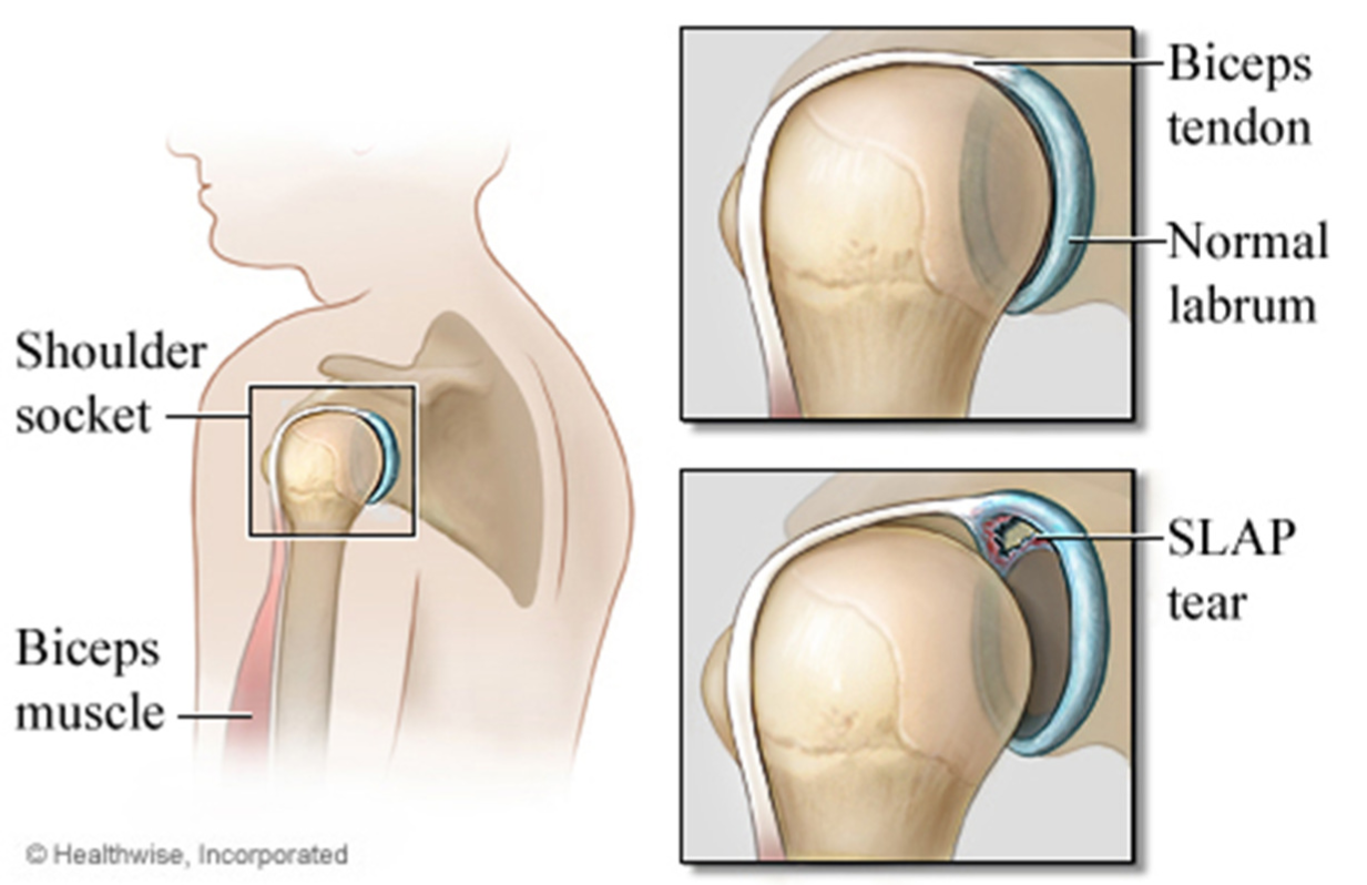
Slap Tear Brisbane Knee And Shoulder Clinic Dr Macgroartybrisbane Knee And Shoulder Clinic from kneeandshoulderclinic.com.au Secondary restaint to inferior translation in the abducted shoulder. The rotator cuff andwhere the bicep tendon meets the shoulder. Normal anatomy, variants and checklist. The tendon of the infraspinatus passes posteriorly on to the. Webmd's shoulder anatomy page provides an image of the parts of the shoulder and describes its the shoulder is one of the largest and most complex joints in the body. Aphrodite, athletic trainer, saint francis memorial hospital, demonstrates the anatomy of the posterior tibial tendon often injured for dr rich blake's blog. The tendons that control movement in your hands, wrists and fingers run through your forearm. Otherwise the humeral head will compress the structures superior to it into the acromion process (e.g.
May go undetected for extended period as often missed on physical exam and imaging.
Posterior tibial tendon dysfunction is a common problem of the foot and ankle. Dr daniel j bell ◉ and dr jeremy jones ◉ et al. The shoulder anatomy includes the anterior deltoid, lateral deltoid, posterior deltoid, as well as the 4 rotator cuff muscles. Upper limb, breast, posterior shoulder, lateral chest wall. • review imaging findings relevant to these causes of pain and discuss a rationale for appropriate use. Visit www.handcare.org for more information about conditions, injuries and treatment of the hand, arm, elbow and shoulder. Secondary restaint to inferior translation in the abducted shoulder. The tendons that control movement in your hands, wrists and fingers run through your forearm. Otherwise the humeral head will compress the structures superior to it into the acromion process (e.g. Infrspinatus tendon and teres minor. Specifically, the four rotator cuff muscles include the following Ligaments are soft tissue structures that connect bones to bones. The rotator cuff andwhere the bicep tendon meets the shoulder.
Upper limb, breast, posterior shoulder, lateral chest wall. Posterior — the back of the shoulder. There are several important ligaments in the shoulder. Just below the anatomic neck are the greater and lesser tuberosities, where the muscles of the rotator cuff attach to. You can see these areas marked with an x in the shoulder anatomy diagram above.
Shoulder Pain Causes Treatment And When To See A Doctor from www.verywellhealth.com Adducts and medially rotates arm; .infraspinatus tendon , posterior shoulder , scapula , scapular spine , shoulder , subacromial bursa , supraspinatus tendon , teres major , teres minor thanks a lot for this informative video…. An image depicting shoulder anatomy can be seen below. Upper limb, breast, posterior shoulder, lateral chest wall. There are several important ligaments in the shoulder. Shoulder tendonitis is the inflammation, irritation and swelling of the tendons in the rotator cuff and bicep. • review imaging findings relevant to these causes of pain and discuss a rationale for appropriate use. Specifically, the four rotator cuff muscles include the following
Being an undergraduate student excites me and inspires me to lean.
.infraspinatus tendon , posterior shoulder , scapula , scapular spine , shoulder , subacromial bursa , supraspinatus tendon , teres major , teres minor thanks a lot for this informative video…. Specifically, the four rotator cuff muscles include the following For more anatomy content please follow us and visit our website: The supraspinatus tendon and subacromial bursa). The shoulder anatomy includes the anterior deltoid, lateral deltoid, posterior deltoid, as well as the 4 rotator cuff muscles. • review imaging findings relevant to these causes of pain and discuss a rationale for appropriate use. The rotator cuff andwhere the bicep tendon meets the shoulder. Upper limb, breast, posterior shoulder, lateral chest wall. Secondary restaint to inferior translation in the abducted shoulder. • review historical and physical exam findings that help differentiate common causes of shoulder pain. The tendons that control movement in your hands, wrists and fingers run through your forearm. Acute tears may occur when the arm is violently pushed into abduction; We hope this picture shoulder tendon muscle bone and nerve anatomy can help you study and research.
Runs along the deltoid tuberosity on the posterior surface of the humerus and contains the radial nerve shoulder tendon anatomy. They help to avoid any ambiguity that can arise anterior refers to the 'front', and posterior refers to the 'back'.
Posterior Shoulder Tendon Anatomy / 4 Muscles And Tendons Of The Rotator Cuff Rotator Cuff Muscle Anatomy Rotator Cuff Injury : Anatomical terms of location are vital to understanding, and using anatomy.. There are any Posterior Shoulder Tendon Anatomy / 4 Muscles And Tendons Of The Rotator Cuff Rotator Cuff Muscle Anatomy Rotator Cuff Injury : Anatomical terms of location are vital to understanding, and using anatomy. in here.

/shoulder_pain_medreview-01-5c3b9f8546e0fb0001bdeaaa.png)

